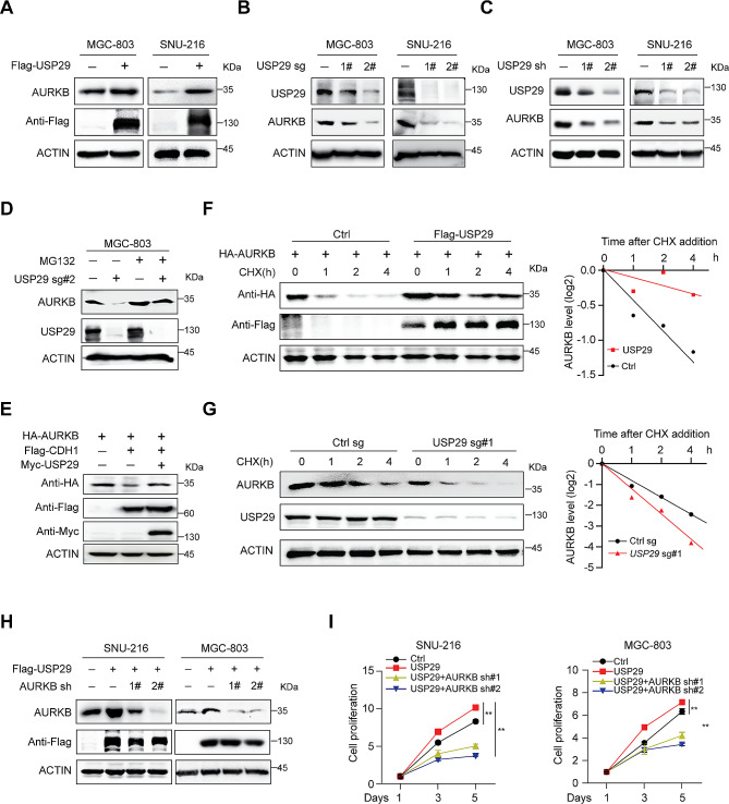
Fig. 3.
USP29 sustains AURKB protein stability. (A) Total cell lysates were extracted from MGC-803 and SNU-216 cells with or without USP29 overexpression, endogenous AURKB level was analyzed by immunoblotting. (B) Cells were infected with control or USP29 sgRNAs, USP29 and AURKB protein expression were detected using immunoblot. (C) Cells were infected with control or USP29 shRNAs, USP29 and AURKB protein expression were detected using immunoblot. (D) MGC-803 cells with or without USP29 depletion were treated with MG132 (10 µM) for 6 h prior to harvest. AURKB and USP29 proteins were analyzed by immunoblot. (E) 293T cells were co-transfected with epitope-tagged AURKB, CDH1 and USP29, immunoblot of respective proteins are shown. The experiments were independently repeated three times with similar results (A–E). (F) 293T cells were transfected with indicated plasmids for 24 h, and treated with 100 µg/mL CHX; protein expression was analyzed with immunoblot and protein quantifications are shown. (G) MGC-803 cells with or without USP29 depletion were treated with 100 µg/mL CHX and harvested at the indicated time, endogenous AURKB protein were detected by immunoblot and protein quantifications are shown. Data shown were obtained from averages of three independent experiments (F, G). (H) Gastric cancer cells were infected with Flag-USP29 and/or AURKB shRNA viruses, the USP29 overexpression and AURKB knockdown were measured by immunoblots. The experiments were independently repeated three times with similar results. (I) Proliferation of stable cell lines in H. Data shown were obtained from mean ± SD of technical triplicates. **p < 0.01

