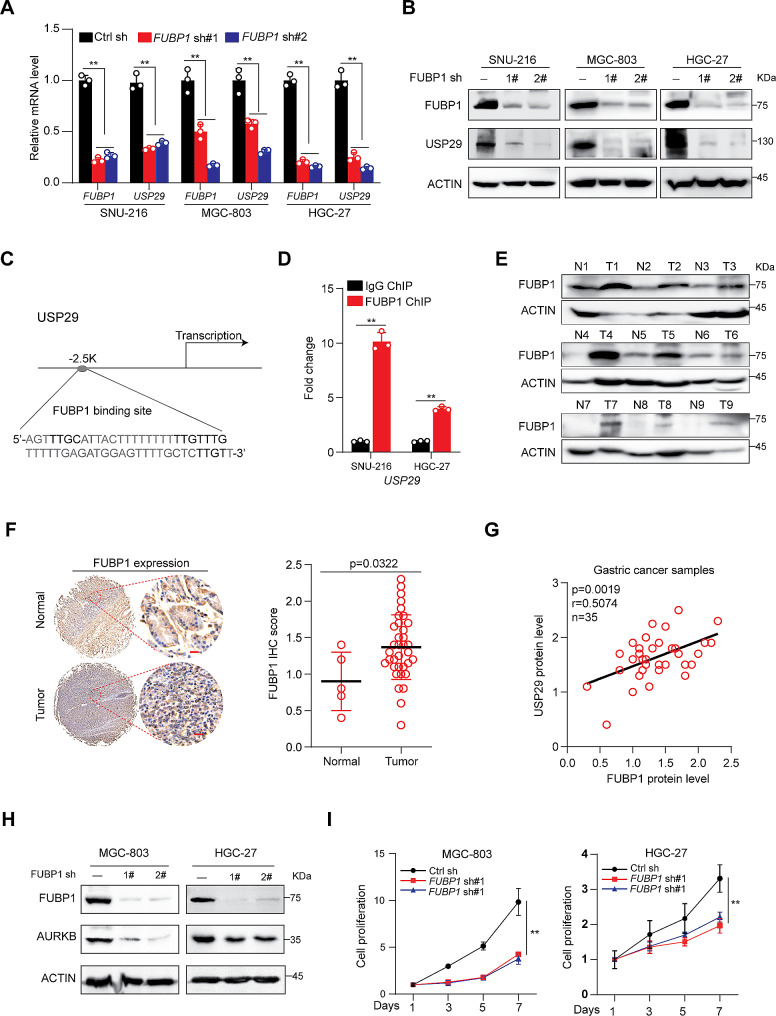
Fig. 4.
FUBP1 overexpression transcriptionally activates USP29 in gastric cancers. (A and B). Gastric cancer cell lines were infected with control or FUBP1 specific shRNAs, the mRNA (A) and protein (B) expression of FUBP1 and USP29 were analyzed using qRT-PCR or immunoblot. Graph shows mean ± SD from triplicates; significance was determined by unpaired two-tailed Student’s t-test (A). The experiments were independently repeated three times with similar results (B). (C). A Schematic presentation of potential FUBP1 binding site on the USP29 promoter. (D). ChIP analysis of FUBP1 binding to the USP29 promoter in SNU-216 and HGC-27 cells. Averages of fold enrichment between the FUBP1 antibodies and IgG were shown. Graph shows mean ± SD from triplicates. (E). FUBP1 protein expression in 9 gastric tumors and paired non-cancerous tissues were analyzed using immunoblot. (F). left: Representative immunohistological images of FUBP1 in 5 normal gastric tissues and 35 tumors. Scale bar: 20 μm; right: quantifications of the immunohistochemistry are shown. (G). Correlation between protein levels of FUBP1 and USP29 in 35 gastric tumors. (H). MGC-803 and HGC-27 cells were infected with control or FUBP1 specific shRNAs, and the expression of FUBP1 and AURKB were analyzed using immunoblots. The experiments were independently repeated three times with similar results. (I). Proliferation of MGC-803 and HGC-27 cells with or without FUBP1 knockdown. Data shown were obtained from mean ± SD of technical triplicates. **p < 0.01

