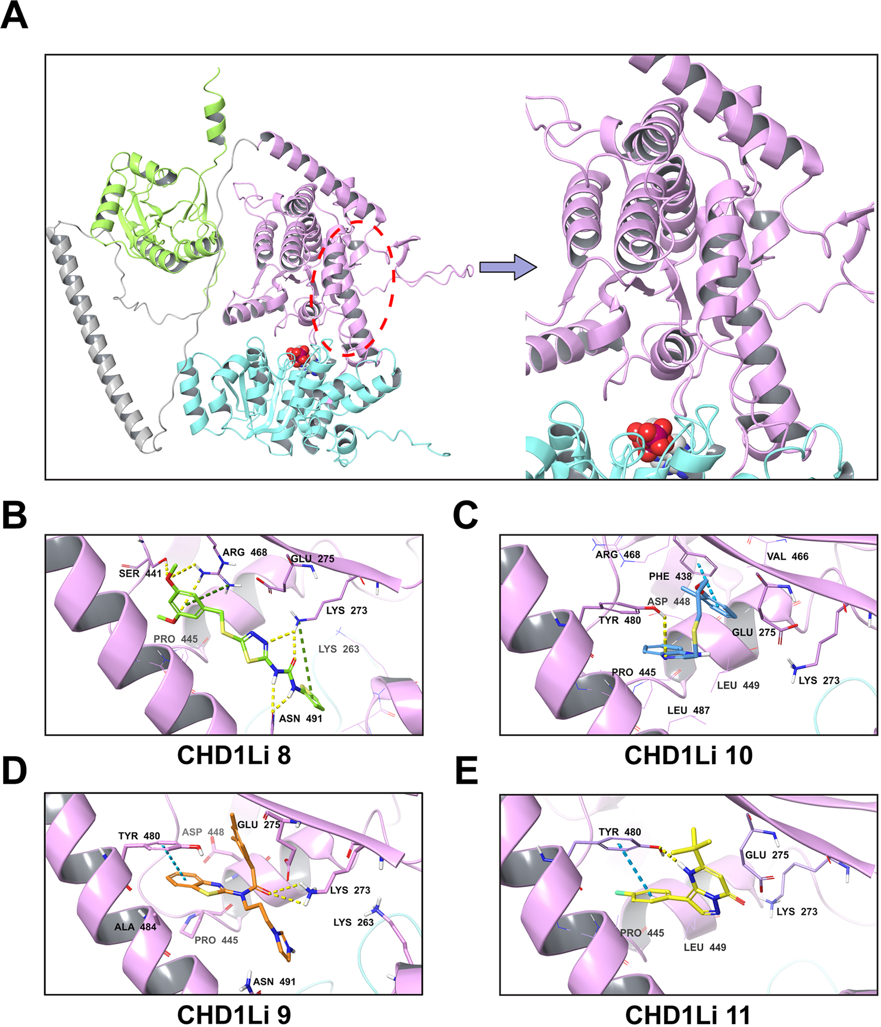
Fig. 5.

Predicted fl-CHD1L allosteric binding site and binding poses of hit CHD1Li. (A) fl-CHD1L structure showing the most plausible CHD1Li binding site. The domain architecture is depicted in cartoon representation as N-ATPase (cyan), C-ATPase (purple), macro domain (green) and linker region (gray). The proposed C-ATPase allosteric binding site is marked with a red ellipse while the ATP binding site is depicted by the bound ADP shown in CPK representation. (B-E). Each panel shows the 3D representation of the predicted binding pose for the CHD1Li as indicated. The non-bonded interactions are depicted as hydrogen bond (yellow-dashed), pi-cation (green-dashed), and pi-pi stacking (blue-dashed).
