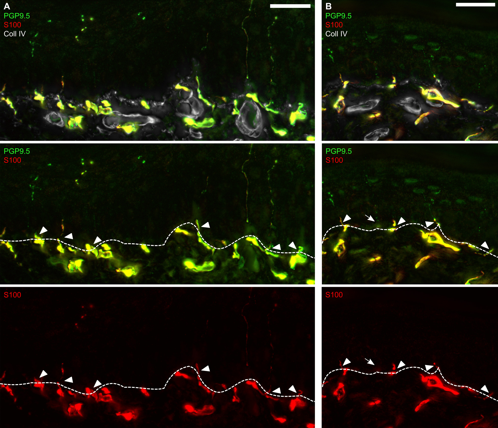
Fig. 1.

Murine intraepidermal Schwann cells in glabrous plantar skin. A, B Representative images of immunostainings of skin sections harvested from hind paw foot pads are displayed (n = 2): Schwann cells were labelled with anti-S100 antibody while intraepidermal nerve fibres were identified with anti-PGP9.5 antibody. The epidermal-dermal border was visualized with anti-Coll IV antibody. Fibres and Schwann cell processes crossing the epidermal-dermal border (dashed line) were counted. Arrows indicate IENF without intraepidermal Schwann cell process, arrowheads intraepidermal Schwann cell processes. Scale bar = 25 µm
