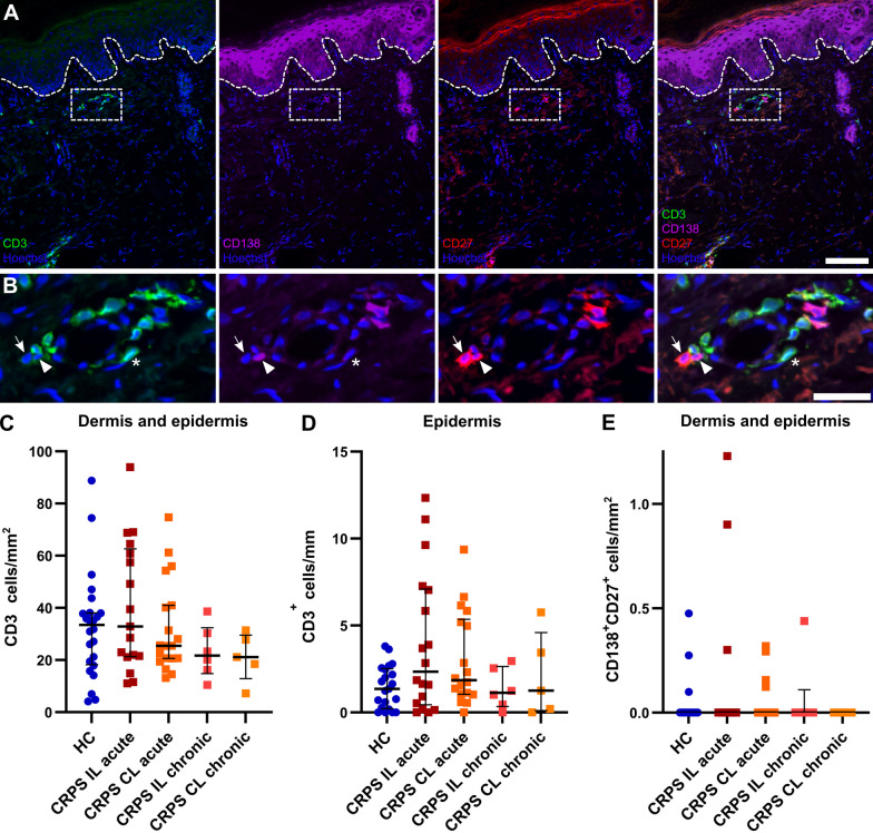
Fig. 6.
No apparent lymphocyte infiltration into skin from patients with acute or chronic CRPS. Skin biopsy of the index finger with A T cells (CD3+; asterisk) and plasma cells (CD27+CD138+; arrow and arrowhead). Scale bar = 100 µm. Dashed box indicates B the zoom-in area with T and plasma cells. Scale bar = 30 µm. C, D Densities of T cells in the dermis and epidermis of acute and chronic CRPS patients and HC are depicted. E Plasma cells (CD27+CD138+) were only detected in 7 samples; outliers were not excluded for plasma cells. Data are shown as median ± interquartile range; Kruskal–Wallis and Dunn's tests; C nHC = 24, nacute IL = 18\1, nacute CL = 19, nchronic IL/CL = 5/6; D nHC = 21\3, nacute IL = 19, nacute CL = 18\1, nchronic IL/CL = 5/6. CL: contralateral; HC: healthy controls; IL: ipsilateral

