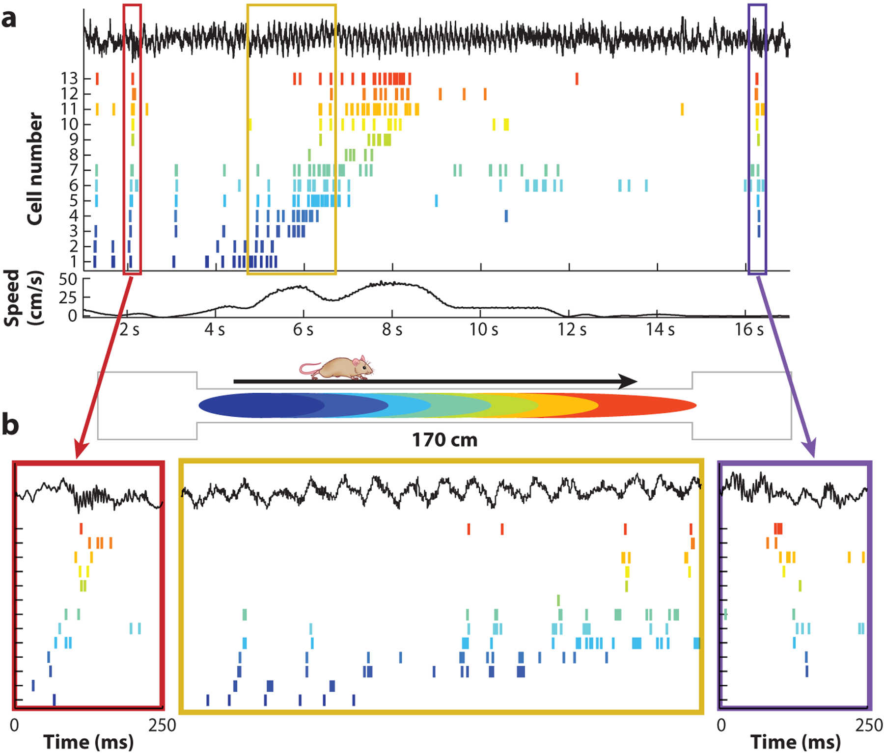
Figure 2.

Time compression of neuronal assembly sequences. (a) Spike trains of 13 hippocampal neurons (color ticks) before, during, and after a single lap. The top black trace illustrates the local field potential; the bottom black line illustrates the locomotion speed of the rat. (b) Spike sequences within single theta cycles are compressed versions of the place field activity on the track (2-s segment highlighted in the yellow box). These theta sequences gradually shift as the animal moves from left to right down the track. On each end of the track (red and purple boxes), spiking during ripples reflects forward and reverse replay of the sequences on the track, respectively. Figure adapted from Diba & Buzsáki (2007).
