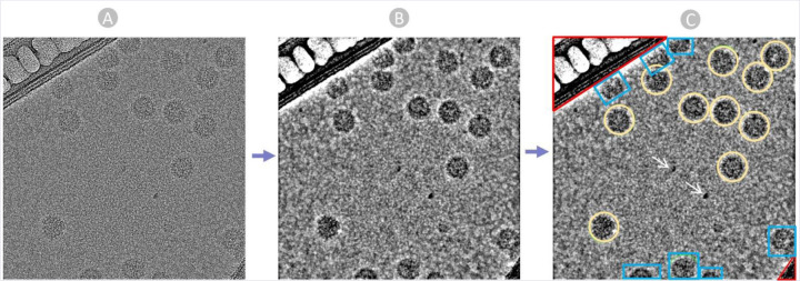
Figure 3:
The manual Picking Process. (A) Raw micrograph obtained from EMPIAR. (B) Preprocessed Micrograph with Low Pass filter: 28 A to ease particle recognition and picking. (C) Manually picked true virus particle encircled in yellow, carbon region colored in Red, ice patches and artifacts pointed by white arrows, and cut particles in edges colored in blue.

