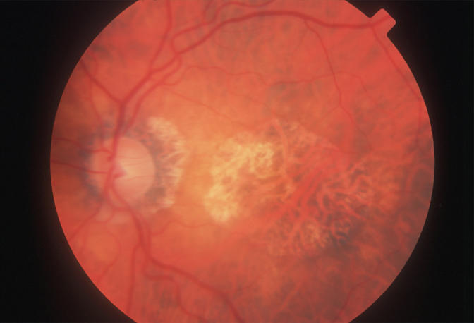
Figure 1.
Fundus photograph showing atrophic or dry age-related macular degeneration. Note the well demarcated area of central retinal, retinal pigment epithelium and choroidal atrophy revealing the pale underlying sclera. Colour version available on [www.jrsm.org]

