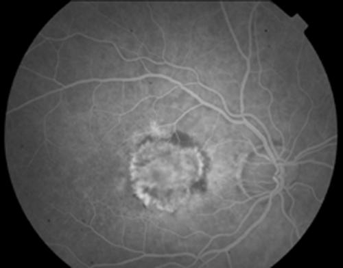
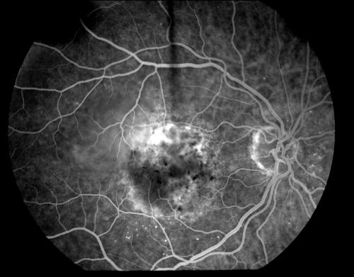
Figure 3.
Fundus fluorescein angiogram showing marked central leakage (bright white area) from a subfoveal lesion classified as classic with occult (a) at presentation and (b) three months after photodynamic therapy. Note that the central area of leakage is much reduced in (b) denoting closure of the choroidal neovascular membrane


