
Fig 2. HLA-C amino acid position in the peptide binding groove associated with rs6906846 A/G (risk/protective).
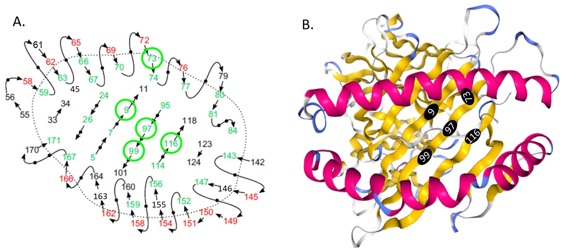
A schematic view of the peptide binding groove and amino acid positions (Adapted from van Deutekom & Keşmir 2015) (A). Residues pointing toward the peptide binding site can interact with the peptide and are colored in green. Residues pointing up from the peptide binding site can interact with the T cell receptor and are colored in red. The remaining residues are shown in black. The 3D structure of the HLA-C molecule (PDB id: 5VGE) and amino acid positions from the Protein Data Bank (PDB: http://www.rcsb.org/3d-view/5VGE) (B). Amino acid positions that are associated with rs6906846 A/G (risk/protective) and pointing towards the peptide binding groove are circled in green (A) and in black filled circles (B).