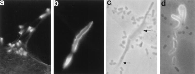
FIG. 4.
Bacteria induce complex extended structures of polymerized actin. HeLa cells were infected with DAEC strain 3431 for 6 h (a) or 3 h (b and c) or with DAEC strain B6 for 3 h (d) and processed as for Fig. 3. Fluorescence (a and b), phase-contrast (c), and simultaneous fluorescence and phase-contrast (d) micrographs are shown. Micrographs b and c are taken from identical areas. Actin accumulates in long horn-like structures protruding from the surfaces of the HeLa cells with a single bacterium on the top (a) or in long tubes associated with bacteria (b to d). Accumulated actin can be detected in phase-contrast micrographs as darker areas (indicated by arrows in panel c). Magnifications: a, ×260; b to d, ×410.

