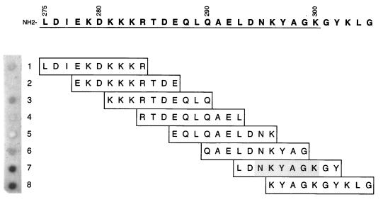
FIG. 3.
Fine mapping of the MAb 10F3-reactive epitope by a dot blot assay involving immobilized synthetic decapeptides. Binding of MAb 10F3 to individual peptides was detected by incubating the membrane with radioiodinated goat anti-mouse immunoglobulin. The autoradiograph of the dot blot (peptides 1 to 8) is shown on the left; the corresponding amino acid sequence of each of the overlapping decapeptides with their respective positions in the CopB protein is listed on the right. The numbers written vertically denote the amino acid positions in the intact CopB protein; the R1 region is underlined.

