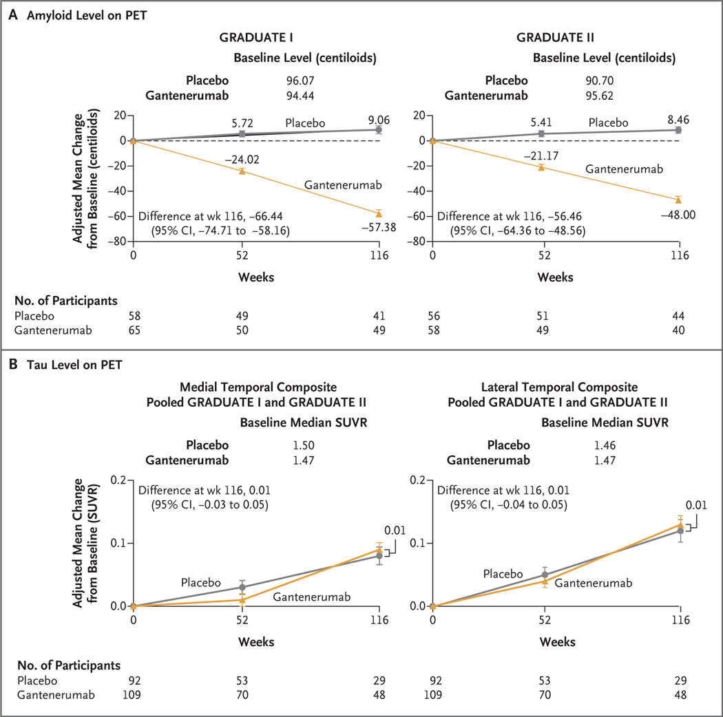
Figure 3. Biomarker Outcomes.
Shown is the adjusted mean change from baseline in the amyloid level (Panel A) and the tau level (Panel B) on positron-emission tomography (PET) through week 116. I bars indicate 95% confidence intervals. In the amyloid PET substudy, the main outcome was the change from baseline to week 116 in the amyloid level. The amyloid level was assessed on florbetaben or flutemetamol PET and was measured as a standardized uptake value ratio (SUVR), which is the ratio of the standardized uptake value in the composite region of interest to the value in the inferior cerebellar cortex; the SUVR results were converted to centiloids. In the tau PET substudy, the main outcome was the change from baseline to week 116 in the tau level. The tau level was assessed in medial temporal, lateral temporal, frontal, and parietal composite regions on PET with 18F-GTP1 (Genentech tau probe 1, an investigational radioligand for in vivo imaging of tau protein aggregates) and was measured as an SUVR.

