. 2024 Jan 17;13:e83725. doi: 10.7554/eLife.83725

Figure 3. Caspase-11 catalytic activity is required for lipopolysaccharide (LPS)-induced oligomerization in HEK293T cells.

(A) HEK293T cells stably expressing 2xFLAG-Casp11 (WT) or 2xFLAG-Casp11 (C254A) were transfected with LPS (1 μg/mL) from S. enterica serovar Minnesota and imaged by fluorescence microscopy 24 hr post-transfection. Casp11 was stained with anti-FLAG-FITC (green), and nuclei (blue) were stained with DAPI. White arrows denote Casp11 specks. Scale bar = 10 μm. (B) Casp11 protein levels in each stable HEK293T cell line were assayed by immunoblotting for FLAG or β-actin (loading control). (C) HEK293T cells stably expressing WT 2xFLAG-Casp11 were transfected with LPS as in (A), in the presence of pan-caspase inhibitor zVAD (200 μM). Casp11 was stained by immunofluorescence using anti-FLAG-FITC (green) and imaged by confocal microscopy (×63 objective). Nuclei (blue) were stained with DAPI. White arrows denote Casp11 specks. Scale bar = 10 μm. Speck formation in (C) was quantified by (D) number of cells per field of view, and (E) number of cells per field of view with at least one speck, denoted as ‘speck formation index.’ Error bars represent mean ± SEM of 14 fields of view (500 cells) from three independent experiments. Data were analyzed by Student’s t-test, ns, not significant, ****p<0.0001.

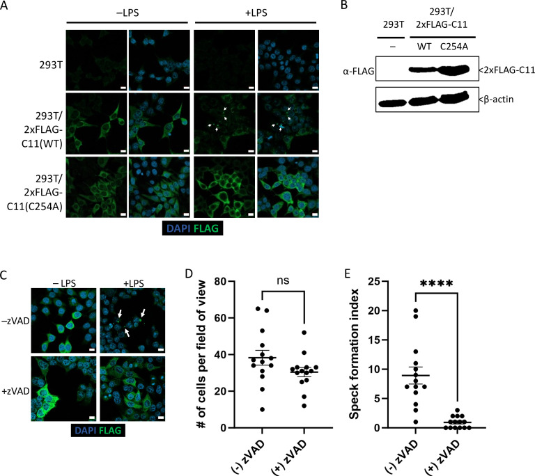
Figure 3—source data 1. Source data for Figure 3B.
2xFLAG-Casp11 protein levels in each stable HEK293T cell line were determined by immunoblotting for FLAG. β-actin was used as loading control.

Figure 3.

Figure 3—figure supplement 1. N- and C-terminal caspase-11 subunits co-localize in caspase-11 specks.

Figure 3—figure supplement 1.

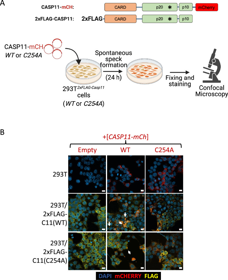
(A) Schematic representing experimental design of colocalization experiment (created with Biorender.com). (B) HEK293T cells stably expressing wild-type (WT) or catalytically inactive (C254A) 2xFLAG-Casp11 were transiently transfected with mCherry-tagged WT or C254A Casp11 DNA constructs for 24 hr. Stably expressed 2xFLAG-Casp11 was stained by immunofluorescence using anti-FLAG (yellow) and cells were imaged by confocal microscopy (×63 objective). Nuclei (blue) are stained with DAPI. White arrows denote Casp11 specks. Scale bars = 10 µM. Images are representative of ≥3 fields of view per condition (150–200 cells) and ≥3 independent biological replicates.
Figure 3—figure supplement 1—source data 1. Source data for Figure 3—figure supplement 1B.
HEK293T cells stably expressing wild-type (WT) or catalytically inactive (C254A) 2xFLAG-Casp11 were transiently transfected with mCherry-tagged WT or C254A Casp11 constructs for 24 hr. Stably expressed 2xFLAG-Casp11 was stained by immunofluorescence using anti-FLAG (yellow) and cells were imaged by confocal microscopy (×63 objective). Nuclei (blue) are stained with DAPI.