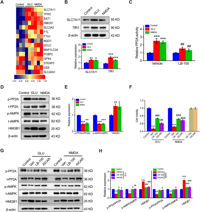
Fig. 2. Activation of SLC7A11 via the PP2A-AMPK axis contributes to NMDAR-activated ferroptosis in HUVECs.
A Differentially expressed genes closely associated with HUVEC ferroptosis in NMDA- or GLU-treated cells determined by RNA-seq. Blue: low expression levels; Red: high expression levels. BProtein blotting and quantitation of SLC7A11 and TfR1 expression in HUVECs in response to GLU or NMDA treatment. *P < 0.05, **P < 0.01 and ***P < 0.001 vs control. C The relative PP2A activity of HUVECs after pretreatment with LB-100 (0.5 μM) followed by NMDA or GLU treatment (n = 6–8). ***P < 0.001 vs control with vehicle; ##P < 0.01 < 0.001 vs control with LB-100. D Protein expression levels and (E) quantitation of SLC7A11 upstream, including PP2A, AMPK, and HMGB1, were analyzed by western blotting (n = 6). *P < 0.05 **P < 0.01, and ***P < 0.001 vs control. F MTT assay detected the cell viability of HUVECs treated with NMDA or GLU and/or LB-100 (0.5 μM) and AICAR (25 μM) for 24 h (n = 6). ***P < 0.001 vs control; ##P < 0.01 and ###P < 0.001 vs GLU; &&&P < 0.001 vs NMDA. G Protein expression levels and (H) quantitation of LB-100 and AICAR with the relative expression of p-PP2A, PP2A, p-AMPK, p-AMPK, and HMGB1 in HUVECs (n = 5). *P < 0.05, **P < 0.01 and ***P < 0.001 vs control; #P < 0.05, ##P < 0.01, and ###P < 0.001 vs GLU; &P < 0.05 and &&&P < 0.001 vs NMDA.

