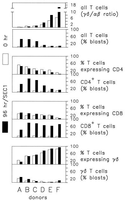
FIG. 1.
Influence of γδ T cells on CD4+ and CD8+ T cells in PBMC cultures following a 96-h incubation with SEC1. Data are percentages of T cells obtained in a representative experiment. Donors A through F were healthy Holstein-Frisian cattle differing in γδ/αβ T-cell ratios as indicated in the figure. Each of the six donors was tested in the following number of replicate experiments: donor A, 7; donor B, 2; donor C, 3; donor D, 4; donor E, 3; and donor F, 6. They consistently yielded similar results.

