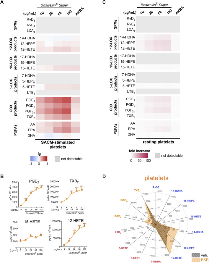
FIGURE 4.
Impact of Boswellin® Super (BSR) on the modulation and induction of LM formation in human platelets. (A,B) 5 × 10⁸ Human platelets were pre-incubated with indicated concentrations of BSR, AKBA (10 µM), or vehicle (0.1% ethanol) for 30 min and then stimulated with 1% SACM for 90 min at 37°C. Formed LM were isolated from the supernatants by SPE and analyzed by UPLC-MS/MS. (A) Heatmap showing the log-fold changes in LM formation for BSR- or AKBA- versus vehicle-pretreated cells stimulated with 1% SACM, n = 3, separate donors. (B) Data are given as pg/5 × 108 cells in line charts (orange) with mean ± S.E.M., n = 3, separate donors. For statistical analysis, data were log-transformed, one-way analysis of variance (ANOVA) with Dunnett’s multiple comparison test, *p < 0.05; ***p < 0.001 against vehicle. (C,D) 5 × 10⁸ Human platelets were incubated with indicated concentrations of BSR, AKBA (10 µM), or vehicle (0.1% ethanol) for 180 min at 37°C. Formed LM were isolated from the supernatants by SPE and analyzed by UPLC-MS/MS. (C) Heatmap showing the fold increase in LM formation for BSR- or AKBA- versus vehicle-treated cells, n = 3, separate donors. (D) Radar plot showing pg of 5 × 10⁶ platelets for selected LM formed by cells after BSR (50 μg/mL) treatment compared to vehicle controls.

