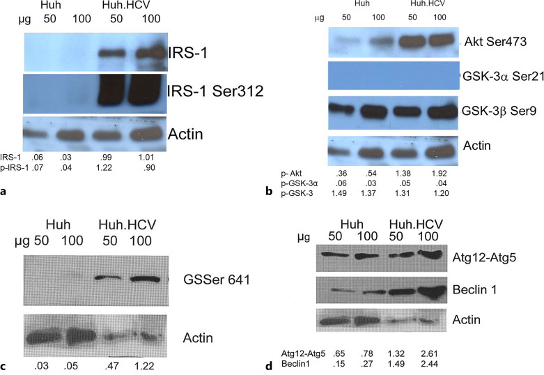
Fig. 1.
Western blot analyses of the key proteins of insulin signaling, glucose homeostasis, and autophagy pathways in acute infection. Huh7 cells were infected with infectious HCV secreted in the media from persistently infected cells (Huh.HCV.2a). Infection was carried out at an HCV genome equivalence (GE) of 5 × 10⁶ and at a GE/cell ratio of 100 (MOI approximately 2.5). Cell lysate was prepared at 48 h for Western blot analyses. Two different amounts of lysate (50 and 100 μg) indicated above each lane were fractionated on a 10% PAGE IRS-1 and IRS-1 Ser312 (a); Akt Ser473, GSK-3α Ser21, GSK-3β Ser9 (b); GS Ser641 (c); autophagy proteins Atg12-Atg5 conjugates and Beclin-1 (d). Actin is the loading control in Western blot. Band intensity was quantified and normalized with respect to this actin control as indicated beneath each lane. When appropriate, further normalization was carried out with respect to uninfected cells.

