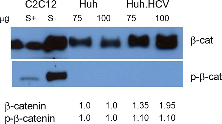
Fig. 4.
Western blot analysis for Wnt/β-catenin (β-cat) using specific antibodies for phosphorylated or nonphosphorylated β-catenin. Lysates from acutely infected cells were prepared and analyzed. Two different amounts of lysates were fractionated (75 μg and 100 μg) as marked above each lane. Lysates from C2C12 muscle cells were induced by serum (S+) or were serum-starved (S−) and used as markers for low- and high-level expression for AMPK and included as controls. Band intensity is indicated beneath each lane. A weak and a strong band were present in the control lanes with an antibody specific for β-catenin and p-β-catenin.

