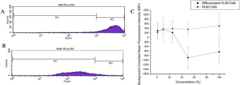
Figure 6.
(A) Flow cytometry data showing undifferentiated HL60 cells incubated with the PE tagged CD71 antibody. (B) Flow cytometry data of differentiated HL60 cells incubated with the PE tagged CD71 antibody. The fluorescence intensity of the differentiated cells shifted to the left indicating the antibody had less transferrin receptors to bind to. Thus, CD71 expression was suppressed (Table S1). (C) Background corrected MFI of the differentiated and undifferentiated HL60 cells stained with transferrin-derived nanoparticles. Statistical difference indicates nanoparticle affinity for CD71 receptor.

