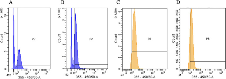
Figure 7.
(A) Flow cytometry data of HL60 cells stained with 3.90 μM affinity CDs. Population 2 (P2) indicated the fluorescence of the affinity CDs and expressed an MFI of 187. (B) HL60 cells incubated in biotinylated CD71 antibody and then stained with 3.0 μM CD71 affinity CDs. Population MFI of 149. The decrease in MFI indicates that the nanoparticles have affinity for CD71. (C) Another population of cells, named population 8 (P8) adjacent to P2, showed a similar shift. The cells stained with 3.90 μM affinity CDs yielded an MFI of 186. (D) However, the cells incubated with the CD71 antibody and then stained with 3.90 μM affinity CDs resulted in a lower MFI of 147.

