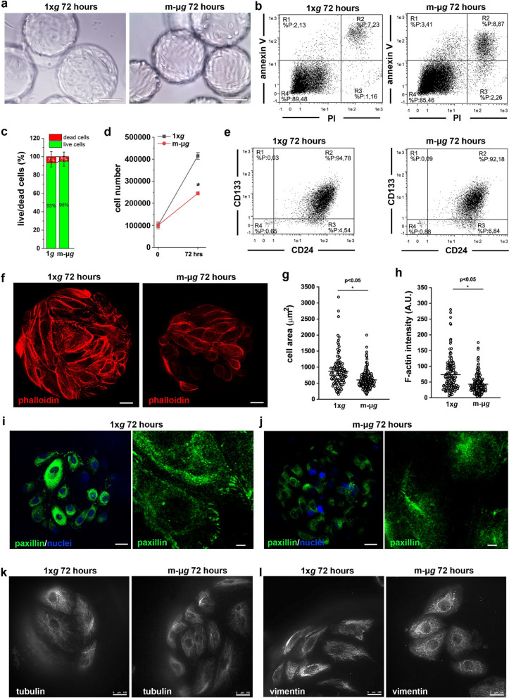
Fig. 2.
Modeled μg conditions reduced proliferation and altered the cytoskeleton of renal progenitor cells. a Phase-contrast images of RPCs grown on Cytodex microbeads at 1 × g (top) and in modeled μg (bottom) conditions. BAR = 100 μm. b Representative flow cytometry analysis of Annexin V/PI staining of RPCs cultured for 72 h at 1 × g (left) and in modeled μg (right) conditions. c Percentage of dead and alive RPCs quantified from confocal images of Calcein-AM/PI staining. Data are expressed as mean ± SEM of two independent experiments. d Growth curves obtained culturing RPCs for 72 h at 1 × g and in modeled μg conditions. Data are expressed as mean ± SEM of three independent experiments. e Representative flow cytometry analysis of CD133 and CD24 staining of RPCs cultured for 72 h at 1 × g (left) and in modeled μg (right) conditions. f Confocal images of phalloidin (red fluorescence) staining in RPCs cultured for 72 h at 1 × g (left) and in modeled μg (right) conditions. BAR = 25 μm. g Measurement of RPC cell area from images as showed in panel (f). At least 50 cells for each condition were analyzed from three independent experiments. h Quantification of phalloidin staining of RPCs cultured for 72 h at 1 × g and in modeled μg conditions. At least 50 cells for each condition were analyzed from three independent experiments. i, j Confocal images of paxillin (green) and nuclei (DAPI, blu) staining in RPCs cultured for 72 h at 1 × g (i) and in modeled μg (j) conditions. BAR = 25 μm and 5 μm, respectively. k Images of tubulin (white fluorescence) staining in RPCs cultured for 72 h at 1 × g (left) and in modeled μg (right) conditions. BAR = 100 μm. l Images of vimentin (white fluorescence) staining in RPCs cultured for 72 h at 1 × g (left) and in modeled μg (right) conditions. BAR = 100 μm. Statistical analysis was performed using the Mann–Whitney test. m-μg: modeled microgravity

