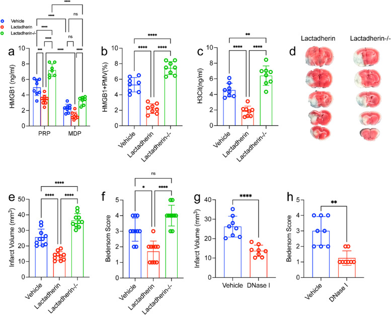
Fig. 6.
Targeting PMVs could regulate NETs formation and brain injury in MCAO model. a PMVs were isolated from lactadherin+/+ or lactadherin-/- mice and incubated for 2 h with neutrophils from WT mice. b Plasma HMGB1 levels were measured by ELISA. c NET releasing cells from each group. Lactadherin+/+ (WT; n = 10) or lactadherin-/-mice or (KO; n = 10) mice were subjected to 1 h of transient middle cerebral artery occlusion followed by 23 h of reperfusion. d NETs were quantified using an H3Cit ELISA from each group. Plasma was isolated and brains were analyzed for ischemic stroke brain damage by TTC staining, 24 h after stroke onset. e Upon TTC staining, live brain tissue will stain red, while dead brain tissue will remain white (outlined with black dotted line). f Infarct size was determined by TTC staining and planimetric analysis. g Neurological score was measured 24 h after stroke using the Bederson Test. Statistics, Brown-Forsythe and Welch’s ANOVA test (g) and ordinary ANOVA (b-d, f). Data are presented as the mean ± SD. *P < 0.05, **P < 0.01, ***P < 0.001 and ****P < 0.0001

