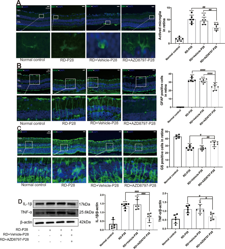
Figure 10.
AZD8797 inhibited the neuroglia activation and inflammatory response. (A) The microglia cells in the RD + AZD8797 group were distributed within the inner retinal layers with branching structure. The number of IBA1 positive cells in the RD + AZD8797 group reduced significantly compared with the RD group. (B) The number of Ly6G-positive cells in the RD group reduced significantly after AZD8797 treatment. (C) The immunostaining intensity of GFAP in the RD group reduced significantly after AZD8797 treatment. (D) The GS immunostaining in the RD + AZD8797 group increased significantly compared with the RD group. (E) Western blot results shown that the IL-1β and TNF-α protein expressions reduced significantly after AZD8797 treatment. (*P < 0.05, **P < 0.01, and ****P < 0.0001, for differences compared with the normal control, and #P < 0.05, ##P < 0.01, ###P < 0.001, and ####P < 0.0001, for differences between animal groups, n = 6).

