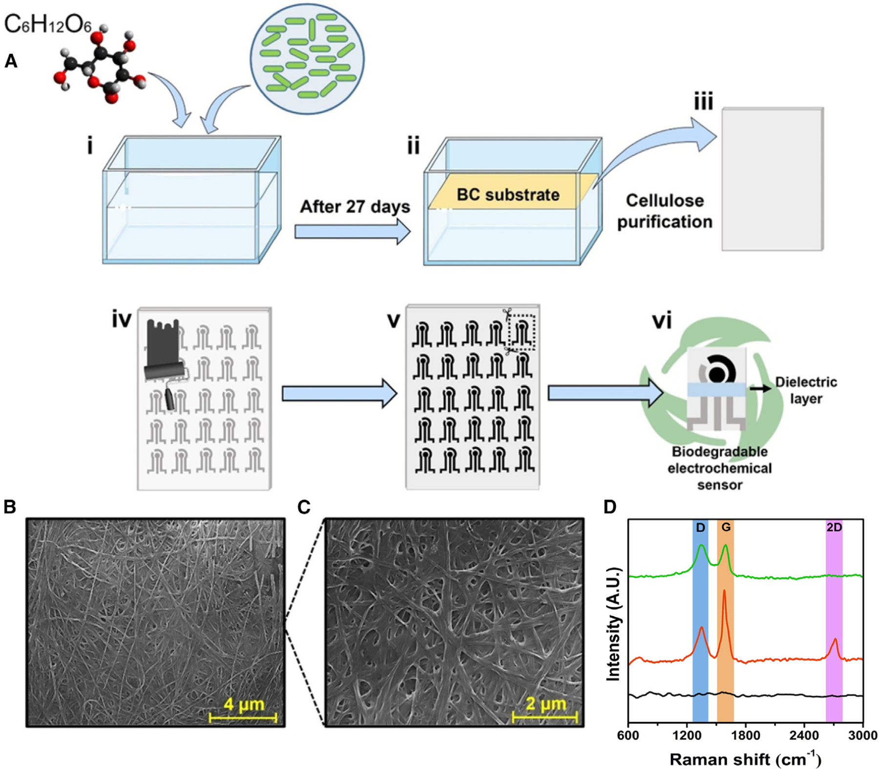
Figure 1. Fabrication and characterization of the SARS-CoV-2 electrochemical biosensor using bacterially produced cellulose.

(A) Fabrication steps of the biodegradable BC substrate and the electrochemical devices. First, the bacterium Gluconacetobacter hansenii was incubated in HS medium with 20 g L−1 glucose (i); after 27 days, a BC substrate was collected and treated with 5 mmol L−1 NaOH at 80°C (ii), resulting in a clear sheet (iii). Next, the biodegradable BC substrate was screen printed with carbon and Ag/AgCl conductive ink (iv), resulting in a device with three electrodes (WE, CE, and RE), which were cut out using a scissor (v), yielding a portable, biodegradable, and inexpensive electrochemical sensor (vi).
(B and C) Micrograph of BC substrate at magnifications of (B) 13,000× (scale bar, 4 μm) and (C) 25,000× (scale bar, 2 μm).
(D) Raman spectra of the (●) BC substrate, (●) BC/carbon ink electrode, and (●) BC/carbon ink/G-PEG electrode. This figure was created in BioRender.com.
