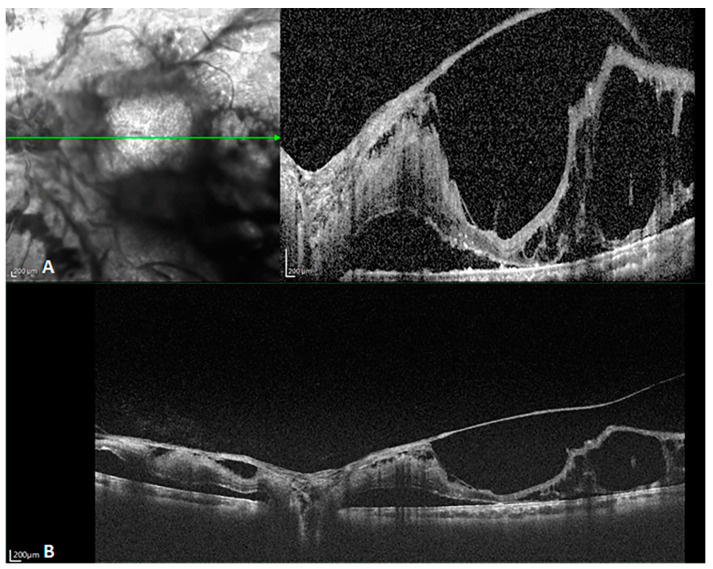
Figure 5.
B-scan images of a patient with proliferative diabetic retinopathy. (A) SD-OCT showing retinal detachment, with the large proliferative membranes, causing a folding artifact. The choroidal layer is incompletely captured. SD-OCT was unable to capture the whole proliferating membrane and the choroidal tissue below because the retinal bulge was too high (The green arrow in the left image indicated the orientation of B scan OCT in the right image). (B) SS-OCT displays an almost identical position. It reveals details of the retina and choroid, even including the choroidal–scleral boundary and part of the sclera. The pre-retinal proliferating membrane extending into the vitreous cavity and the retinal detachment on the nasal side of the optic disk are also clearly visible.

