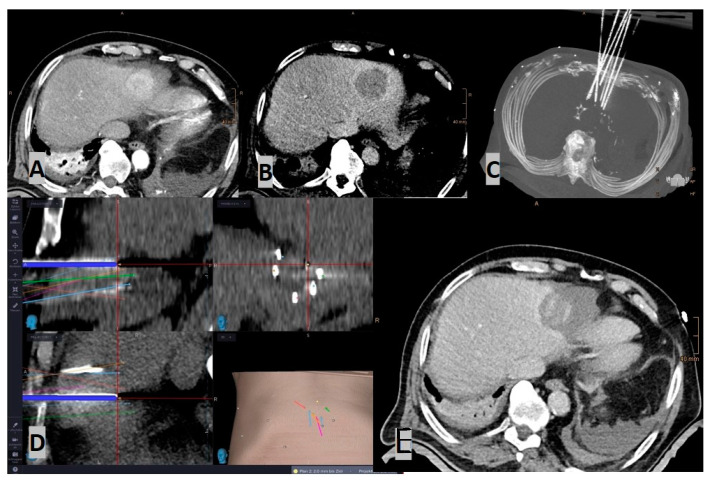
Figure 2.
Case of a stereotactic radiofrequency ablation (SRFA) in an 85-year-old male with a sub-cardiac HCC (4.8 cm): (A) Arterial phase planning CT; (B) Portal-venous phase planning CT; (C) MIP of the control CT, showing in total 7 inserted coaxial needles; (D) Screenshot of the frameless stereotactic navigation system: Superposition of the needle control CT on the planning CT, with the pathways showing precise placement of all needles; (E) Contrast-enhanced control CT (portalvenous phase), showing the large ablation zone covering the HCC, including a sufficient safety margin, which was confirmed by image fusion.

