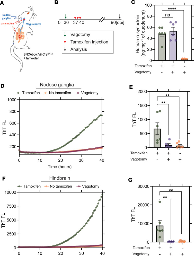
Figure 5. Vagotomy spares the nodose ganglia from α-synuclein seeding activity and prevents spread to the hindbrain.
(A and B) Experimental model. SNCAbow Vil-CreERT2 mice underwent bilateral subdiaphragmatic vagotomy or sham surgery 1 week before tamoxifen treatment. (C) ELISA measurements of α-synuclein protein in the gut 3 months after tamoxifen treatment. RT-QuIC analysis of (D and E) vagal nodose ganglia and (F and G) hindbrain analyzed 3 months after tamoxifen treatment. Representative ThT fluorescence profiles are shown in D and F. All the group analyses are shown as mean ± SEM. All RT-QuIC curves shown are representative of the mean from all the groups analyzed. Data points in B, E, and G represent the significance determined by a 1-way ANOVA with a Dunnett’s post hoc analysis relative to tamoxifen-treated and no-vagotomy groups. **P < 0.01, ****P < 0.0001, n = 6.

