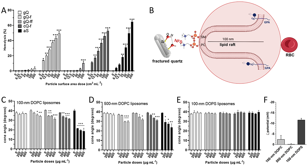
Fig. 2.

Red blood cell membrane lysis and cone angle deformation of DOPC and DOPS liposomes induced by different silica particles. (A) Hemolytic activity of the silica particles incubated with red blood cells. Hemoglobin released from RBCs treated with increasing surface area doses (0, 6.25, 12.5, 25, 50, 100, and 200 cm2 mL−1) of gQ, gQ-f, gQ-ff, cQ-f (positive reference quartz), and aS for 30 min. Data are the mean ± SEM of three independent experiments and were analyzed with one-way ANOVA, followed by Dunnett’s post-hoc test. *p < 0.05, * *p < 0.01, * **p < 0.001 vs. control not exposed to silica particles. (B) Schematic representation of the RBC membrane structure evidencing the major syaloglycoprotein (Glycophorin A, GPA) that confers negative charge to the outer membrane; a sphingomyelin-enriched lipid protrusion deprived of GPA, where SM and PC are evidenced. Phosphocholine polarheads might interact with NFS on silica. (C) Cone angle deformation of liposomes incubated with the silica particles. Cone angle deformation of (C) 100 nm DOPC, (D) 500 nm DOPC, and (E) 100 nm DOPS liposomes treated with gQ’ gQ-f, gQ-ff, cQ-f, and aS for 2 h. Data are the mean ± SEM of three independent experiments and were analyzed with one-way ANOVA, followed by Dunnett’s post-hoc test. *p < 0.05, * *p < 0.01, * **p < 0.001 vs. control not exposed to silica particles. (F) ζ potential measured by ELS in experimental medium at pH 7.4.
