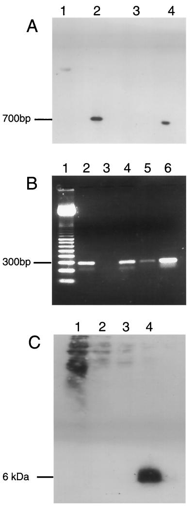
FIG. 6.
(A) Southern blot of EcoRI-digested plasmid and purified viral DNA. Lanes: 1, nonrecombinant insertion plasmid, pSC11; 2, pSC11-ESAT-6; 3, rVV–β-galactosidase; 4, rVV–ESAT-6. (B) RT-PCR of virally encoded ESAT-6 transcripts. Lanes: 1, 100-bp pair molecular mass marker; 2, DNase-treated, RT step included; 3: DNase treated, no RT step; 4, no DNase, no RT step (contaminating viral DNA); 5, no DNase, no RT step; 6, plasmid containing esat-6 gene (positive control). (C) Western blot of rVV-infected cell lysates in a large-gel system with 285 μl of cell lysate/well. Lanes: 1, neat cell lysate; 2, diluted 1:2; 3, 1:4; 4, M. tuberculosis culture filtrate (positive control).

