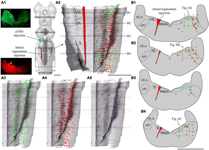
FIGURE 5.
Localization of neurons in the caudal hindbrain (OLA excluded) projecting to the pTRG and the lateral tegmentum on the other side: both injections were made on the same side of the brain. (A1) Schematic drawing of a dorsal view of the whole lamprey brain indicating the location of the injection sites. To the left, photomicrographs illustrating the injection sites on transverse sections for the animal represented in the figure. (A2) 3D rendering of the caudal hindbrain with neurons projecting exclusively to the pTRG in green, exclusively to the lateral tegmentum on the other side in red, and projecting to both in blue. The location of the respiratory VII, IX, and X motor nuclei is indicated by dashed lines. (A3) Representation of the neurons that project exclusively to the pTRG as they appear in panel (A2). (A4) Representation of the neurons that project exclusively to the lateral tegmentum on the other side as they appear in panel (A2). (A5) Representation of the neurons that project to both the contralateral pTRG and contralateral tegmentum as they appear in panel (A2). (B1–B4) Drawings of transverse sections of the caudal hindbrain at the levels indicated on the 3D rendering. The color code is the same as in A. IX, glossopharyngeal motor nucleus; MRRN, middle rhombencephalic reticular nucleus; OLA, octavolateral area; PRRN, posterior rhombencephalic reticular nucleus; pTRG, paratrigeminal respiratory group; rdV, descending root of the trigeminal nerve; VII, facial motor nucleus; X, vagal motor nucleus. Scale bars = 1 mm.

