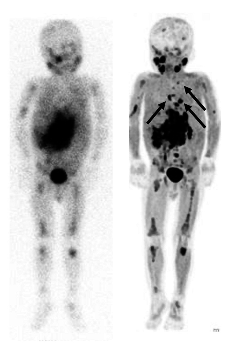
Fig. 2.

Therapy response evaluation of a 4-year-old boy with high-risk neuroblastoma. [123I]mIBG whole-body scan (left panel). Maximum intensity projection (MIP) of [.18F]mFBG PET (right panel). Pathological uptake in primary abdominal neuroblastoma and extensive osteomedullary neuroblastoma localisations on both investigations, more clearly and with higher resolution depicted with [18F]mFBG PET. [18F]mFBG PET also detected additional mediastinal lymph node metastases (arrows)
