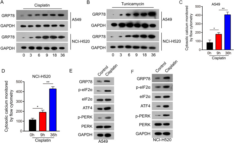
Fig. 3. Cisplatin evoked ER stress in NSCLC cells.
A GRP78 expression was examined in A549 and NCI-H520 cells following treatment with cisplatin (5 μmol/L). Floating and adherent cells were lysed at 0, 3, 6, 9, 18, and 36 h and analyzed by western blot analysis. B GRP78 expression was examined in A549 and NCI-H520 cells following treatment with tunicamycin (200 ng/μl). Floating and adherent cells were lysed at 0, 3, 6, 9, 18, and 36 h and analyzed by western blot analysis. C, D Treated with 5 µmol/L cisplatin for 0, 9, and 36 h, A549 cells (C) and NCI-H520 cells (D) were loaded with Fluo-3am for 30 min. Cytoplasmic calcium level was examined by flow cytometry assay. Data are expressed as mean ± standard deviation. E, F Molecular markers of the endoplasmic reticulum stress signaling pathway were detected by western blot analysis in A549 and NCI-H520. Data are shown as the means of three independent experiments or representative data. Data are expressed as mean ± standard deviation. *P < 0.05, **P < 0.01 by Student’s t test.

