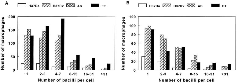
FIG. 2.
Frequency distribution of the numbers of bacilli in macrophages infected with M. tuberculosis. Monocyte-derived macrophages were cultured with H37Ra, H37Rv, and clinical isolates AS and ET for 10 days. Acid-fast staining was performed, and approximately 10,000 macrophages were counted in each experiment. Panels A and B each represent a separate experiment and show the numbers of macrophages containing the indicated numbers of bacilli per cell after 10 days.

