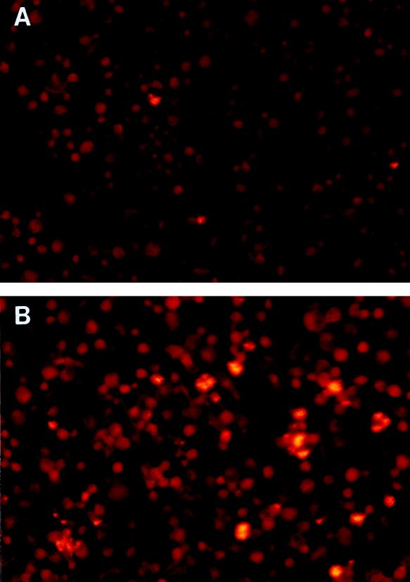
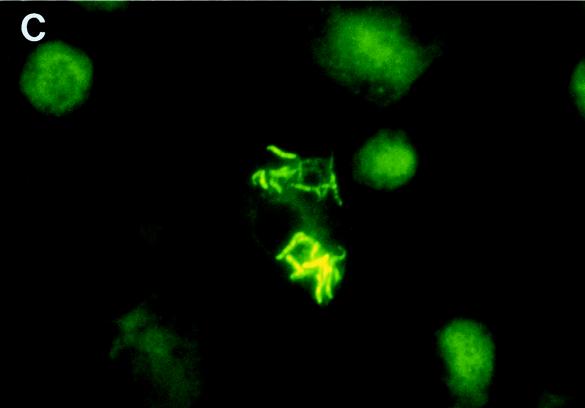
FIG. 3.
Photomicrographs of M. tuberculosis H37Ra (A) and H37Rv (B and C) after 10 days of culture in monocyte-derived macrophages. Panels A and B are low-power-magnification views (×100) of M. tuberculosis cells in macrophages after staining with auramine-rhodamine. The photomicrographs were taken with a fluorescence microscope equipped with a 470- to 490-nm filter (U-MNB; Olympus, Tokyo, Japan). Bacilli are bright yellow, and macrophages are dull red. Panel C shows a high-power-magnification view (×1,000) of M. tuberculosis H37Rv in macrophages after staining with auramine-rhodamine. A 510- to 550-nm filter (U-MWG; Olympus) was used to view the slide. The bacilli are bright yellow, and the macrophages are green. A different filter was used for panel C because it permitted a more distinct view of cell and bacillary outlines at high magnification.


