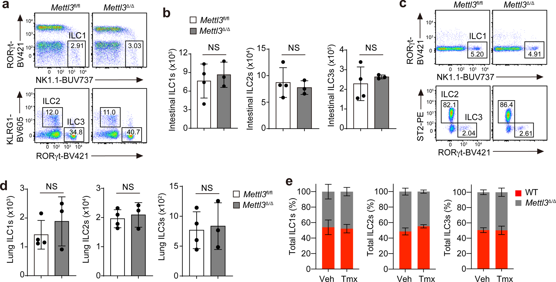
Fig. 1. m6A is dispensable for mature ILC maintenance at steady state.

a,b, Representative flow cytometry (a) and quantification (b) of intestinal ILCs pregated as live CD45+Lin−Thy1+ and further gated as RORγt−NK1.1+ ILC1s, KLRG1+ ILC2s and RORγt+ ILC3s on day 5 after the last tamoxifen (Tmx) dose in Mettl3Δ/Δ mice or Mettl3fl/fl littermate controls treated i.p. with tamoxifen every other day for three doses; not significant (NS) P = 0.6189 (left), NS P = 0.6149 (middle), NS P = 0.5229 (right; b). c,d, Representative flow cytometry (c) and quantification (d) of lung ILCs in mice as in a; NS P = 0.4020 (left), NS P = 0.6529 (middle), NS P = 0.8045 (right; d). e, Ratios of wild-type B6.SJL (WT) and Mettl3Δ/Δ ILCs on day 60 after a mixed transfer of total wild-type and Mettl3Δ/Δ BM cells at a ratio of 1:1 into Rag2−/−Il2rg−/− recipient mice that received three doses of tamoxifen from days 28 to 32 after transfer. Veh, vehicle. Data were analyzed by unpaired two-tailed t-test; n = 3 or 4 in each group in b, d and e. Data are shown as mean ± s.d. Results are representative of three independent experiments.
