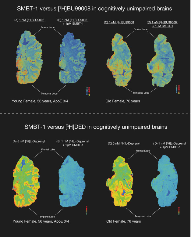
Figure 4.
SMBT-1 versus [3H] l-deprenyl and [3H]-BU99008 autoradiography on large frozen postmortem CN brain sections. The autoradiograms in the figure show the total binding of 5 nM [3H]-l-deprenyl and 1 nM [3H]-BU99008 in young and old CN brains. Coincubation with 1 μM non-radiolabeled SMBT-1 completely displaced [3H]-l-deprenyl binding but only partially displaced [3H]-BU99008 binding from CN brains. We performed semiquantitative analyses of manually drawn ROI to calculate the total binding values (in fmol/mg) and the % of SMBT-1 displacement. Frontal and temporal lobe regions are marked with dark black bars. Results are presented in Table 2. The autoradiography images were adjusted to standardize the color/threshold levels for comparison (standards: 0.84–2960 fmol/mg). For example, autoradiograms of 1 nM [3H]-BU99008 alone and in the presence 1 μM non-radiolabeled SMBT-1 were at the same level. CN, cognitively normal; ROI, region of interest.

