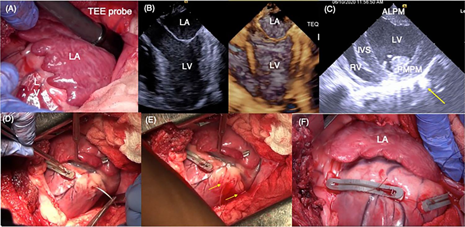
Fig. 2.

A Intra-operative view of the left atrium and the basal aspect of the left ventricle through a left thoracotomy. The transesophageal echo probe was tunneled onto the roof of the left atrium. B 2D and 3D echocardiographic views of the left atrium, mitral valve, and the left ventricle are shown. The papillary muscle position is identified in this view by palpating the epicardium. C Short-axis view of the left ventricle depicting the two papillary muscles in the left ventricle. Epicardial vascular clips were placed at regions where the papillary muscles were identified upon palpation. Yellow arrow indicates reflection from a clip on the epicardium. D Representative photograph depicting placement of the posterior snare, by inserting the needle through the myocardium on the lateral side of the papillary muscle and externalizing on the medial side. E Two ends of the suture externalized through the myocardium, encircling the chordae of the posteromedial papillary muscle in the left ventricle. F Free ends of the snares were tethered maximally and then tied onto a polymer bridge that lifted the snares off the epicardium to avoid coronary compression
