
Fig. 4.
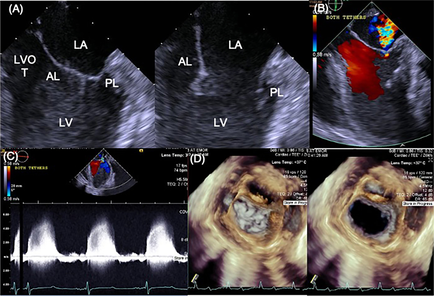
A 2D B-mode view of the mitral valve in systole and diastole with the transesophageal probe placed on the roof of the left atrium. B A representative image of mitral regurgitation in this same view. C Continuous wave imaging of the mitral regurgitation, without the need for angle correction, providing accurate assessment of the severity of regurgitation. D 3D views of the mitral valve from the same probe position