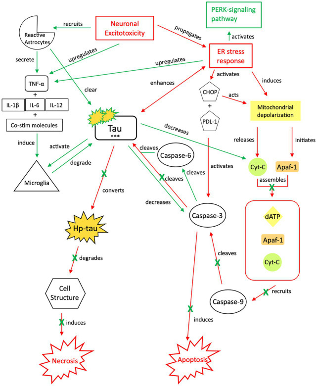
Figure 4.
Our proposed neuroprotective response mechanism involving tau (NPT). Neuronal excitotoxicity imbalances the ER stress response, which activates two pathways: the PERK pathway, responsible for reverting the cell to homeostasis and preserving its integrity, and pro-cell death signaling cascades via CHOP and rapid mitochondrial depolarization, such as apoptosis. In typical apoptotic signaling, mitochondrial depolarization initiates Apaf-1 and releases cyt-c. Cyt-c, with Apaf-1 and dATP, assembles into an apoptosome complex (119–121). The apoptosome complex recruits caspase-9, caspase-9 cleaves caspase-3, and caspase-3 activates apoptosis (122, 123). Tau preserves cellular integrity and reverts cellular signaling away from pro-cell death signaling cascades. Although reduction of caspase-3 cleavage of tau reverts the cell away from apoptotic signaling, tau is cleaved by additional caspases such as caspase-6, resulting in tau phosphorylation (124–128). The increased presence of p-tau decreases the concentration of cyt-c and caspase-3, thereby further inhibiting apoptotic signaling (122, 129, 130). To avoid additional cell death pathways (i.e., necrosis), increases in cytokine expression, TNF-α, and tau concentrations recruit reactive astrocytes and microglia to break down excess tau into non-toxic components (131–136). Successful breakdown of accumulated tau by microglia and reactive astrocytes downregulates pro-death signaling pathways and restores cellular homeostasis. PERK, protein kinase R-like ER kinase; TNF, tumor necrosis factor; IL, interleukin; Co-stim, co-stimulatory (molecules); cyt-c, cytochrome-c; Apaf-1, apoptotic peptidase activating factor-1; dATP, deoxyadenosine triphosphate; NFTs, neurofibrillary tangles; Red, Pro-death signaling; Green, Neuroprotective signaling; o-tau, tau oligomers; t-tau, total tau; p-tau, phosphorylated tau; ***, O-tau, t-tau, p-tau; X, response reduction/down-regulation.

