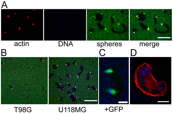
Figure 1.
Appearance of cells migrating on fluorescent microspheres. (A) F-actin (red), DNA (blue), microspheres (green), and merged view indicate that cells clear non-fluorescent tracks in the dense particle field as they move. Bar, 100 μm. (B) Cell lines exhibit differences in motility reflected by the area of particles cleared, highlighted by comparing T98G and U118MG glioblastoma lines. Bar, 300 μm. (C) Cells transfected with expression vector for GFP can be visualized on a blue fluorescent microsphere field. The trails from both cells converge at a common origin, suggesting that the two cells arose from the division of a common progenitor and migrated away. Bar, 20 μm. (D) Confocal section of phalloidin-stained U118MG cell migrating on field of blue microspheres. Bar, 20 μm.

