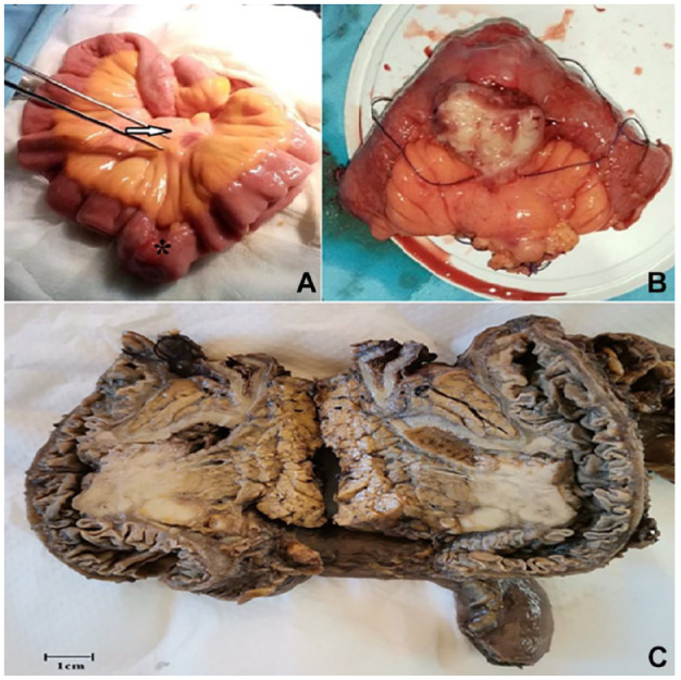
Figure 2.

(A) Intraoperative views showing the tumor (arrow) with mesenteric lymphadenopathy (asterisk). (B) Gross examination revealed a polypoid mass within the small intestine wall. (C) On the cut section, the mass had a tan-white lobulated appearance.
