
Figure 3.
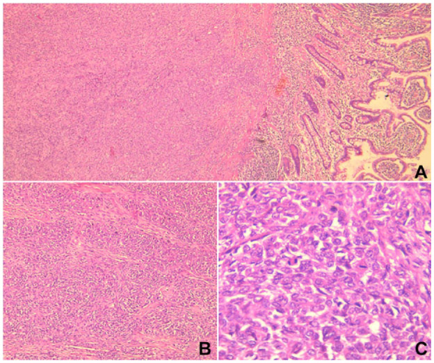
(A) Microscopic examination revealed an infiltrative proliferation centered in the submucosa of the ileum (H&E stain, ×40). (B) The proliferation displayed a nested pattern of growth (H&E stain, ×100). (C) The tumor was composed of uniform epithelioid and spindle cells with variable amounts of eosinophilic cytoplasm. The nuclei were vesicular with inconspicuous nucleoli and mitotic figures. (H&E stain, ×400).
Abbreviation: H&E, hematoxylin and eosin.