Figure 4.

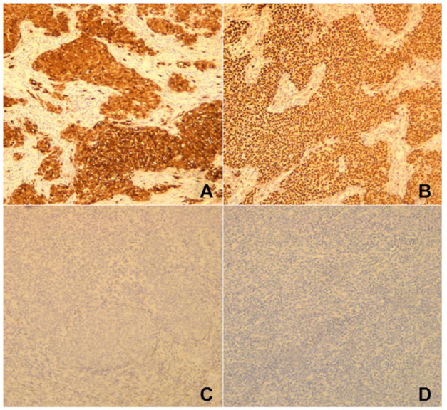
(A) Diffuse and strong staining with S100. (B) Diffuse and strong staining with SOX10. (C) Staining with HMB45 was negative. (D) The tumor cells did not express Melan A.

(A) Diffuse and strong staining with S100. (B) Diffuse and strong staining with SOX10. (C) Staining with HMB45 was negative. (D) The tumor cells did not express Melan A.