
Figure 4.
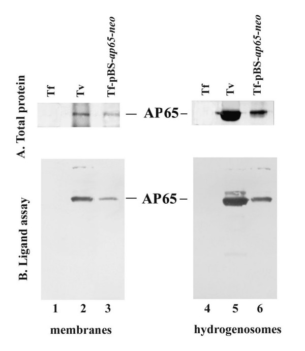
Immunoblots with specific mAb 12G4 detecting AP65 immunoprecipitated from extracts of total trichomonad protein preparations (A) and of proteins after the ligand assay (B) derived from purified hydrogenosomes (lanes 1 through 3) and membrane fractions (lanes 4 through 6). AP65 was immunoprecipitated with mAb from protein extracts from hydrogenosomes and membrane fractions of T. foetus (Tf; lanes 1 and 4), T. vaginalis (Tv; lanes 2 and 5), and transfected T. foetus (Tf-pBS-ap65-neo; lanes 3 and 6). Immunoprecipitated AP65 was prepared as described in the Experimental design section and used in a ligand assay to monitor the amount of AP65 adhesin bound to MS-74 VECs as shown above in Figure 3.