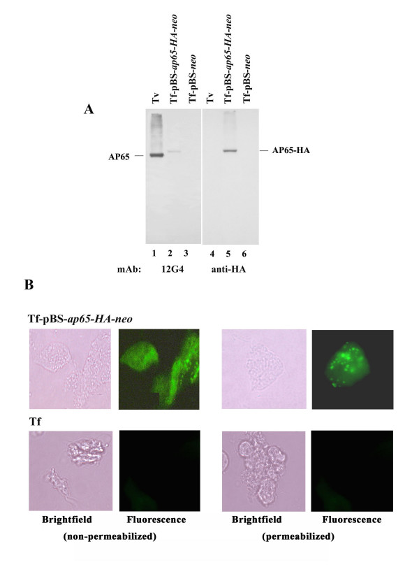
Figure 6.
Immunoblot detection of AP65-HA in T. foetus transfected with pBS-ap65-HA-neo, and immunofluorescence microscopy detecting AP65-HA protein on the surface (non-permeabilized) and in hydrogenosomes (permeabilized). (A) Duplicate immunoblots prepared from total proteins as above were probed with 12G4 mAb to AP65 (lanes 1 through 3) and anti-HA mAb (lanes 4 through 6). AP65 was readily detected by 12G4 in blots of T. vaginalis (lane 1) and T. foetus transfected with pBS-ap65-HA-neo (lane 2) but not T. foetus with control plasmid (lane 3). The anti-HA mAb detected the AP65-HA protein only in T. foetus transfected with pBS-ap65-HA-neo (lane 5). No protein was detectable by anti-HA mAb in blots of control T. vaginalis (lane 4) and T. foetus with the pBS-neo plasmid (lane 6). As an additional negative control for all immunoblots, no protein was detected in the absence of primary antibody or using control hybridoma supernatant. (B) Fluorescence is visualized using anti-HA mAb only in the T. foetus organisms transfected with the pBS-ap65-HA-neo plasmid but not the pBS-neo plasmid. Not unexpectedly, no fluorescence was seen using anti-HA mAb with T. foetus and with the use of control hybridoma supernatant lacking mAb to AP65. Finally, no fluorescence with anti-HA mAb was obtained using T. vaginalis organisms under identical experimental conditions.

