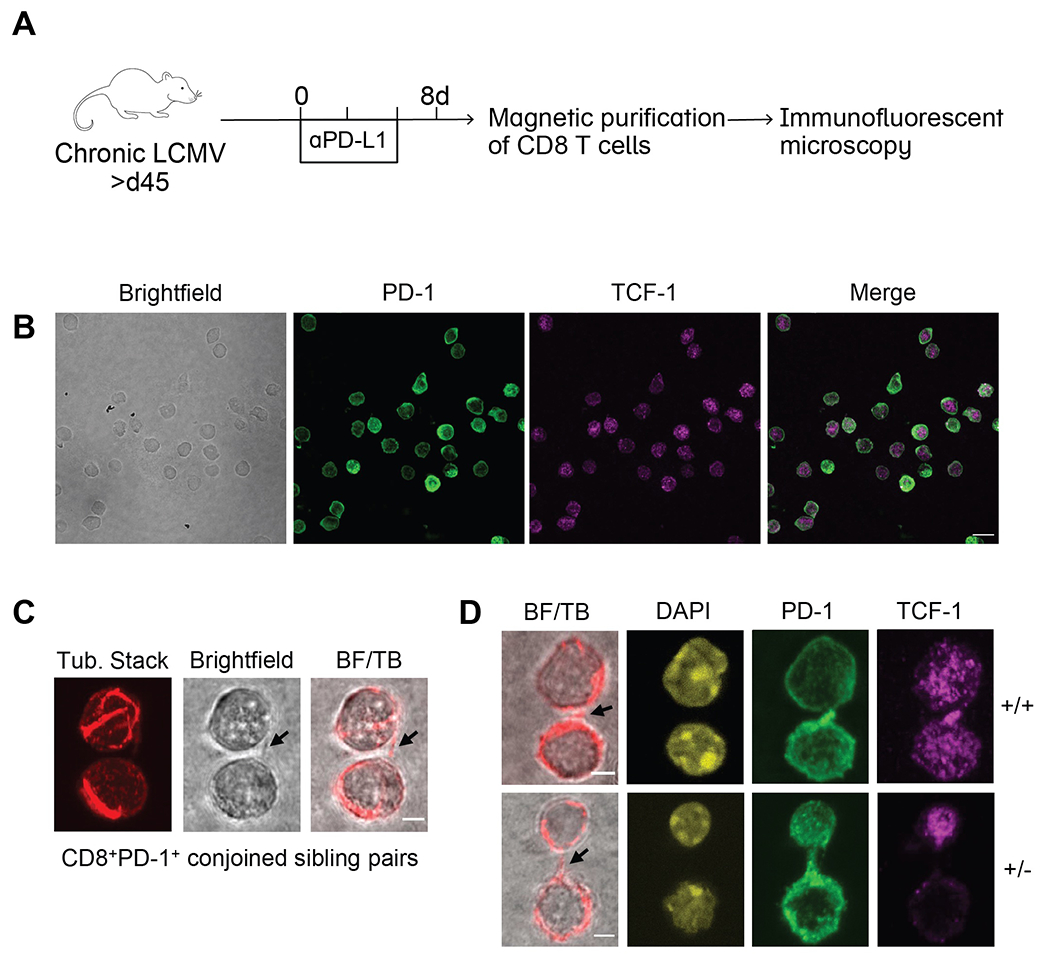
Fig. 5. Asymmetric division can sustain the stem-like CD8 T cell population.

(A) LCMV chronically infected mice were treated with or without αPD-L1 every 3 days. CD8 T cells were isolated from spleens by magnetic purification on day 8 and immunofluorescence confocal microscopy was performed. (B) Representative photos of singlet CD8 T cells following αPD-L1 treatment were captured on 60X lens with 2X scanning zoom (0.1μm/pixel). Merged fluorescence of PD-1 and TCF-1 are shown. Scale bar is 10μm. (C) Conjoined CD8+PD-1+ sibling pairs were captured on 60X lens with 4X scanning zoom (0.05 μm/pixel). Representative conjoined sibling cell pair illustrating bridge between sibling cells marked by arrows in (l-to-r) brightfield and merge of brightfield with single z-slice tubulin (BF/TB). (D) CD8+PD-1+ sibling pairs from αPD-L1-treated mice illustrating TCF-1 concordant (+/+; top) and TCF-1 discordant (+/−; bottom) divisions. (−/−) sibling pairs were also observed but are not shown. Representative images displaying (l-to-r), brightfield merge with single z-slice tubulin (bridges marked by arrows), DAPI, PD-1, and TCF-1 staining (n=14 doublets imaged per group). Scale bars are 2.5μm. (B) and (C) each show representative images from a total of 3 independent experiments.
