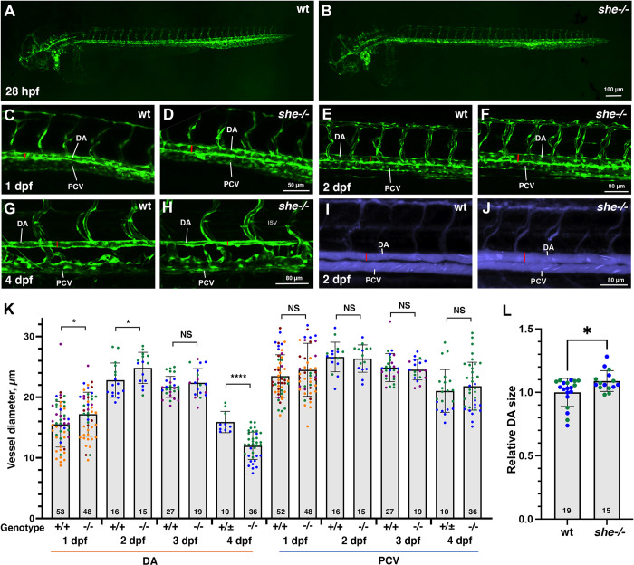
Fig 2. she mutants show enlarged diameter of the dorsal aorta.
(A,B) Overall vascular patterning of she-/- mutants is normal when compared to their sibling wild-type embryos at 28 hpf. Embryos are in kdrl:GFP background. (C-F) A wider DA is observed in she mutant embryos compared to their wild-type (she+/+) siblings at 1 and 2 dpf (28 and 48 hpf respectively). Red line indicates DA diameter. (G,H) DA is narrower in she mutants at 4 dpf compared to their siblings. (I,J) Qtracker dots were injected into the circulatory system at 2 dpf (48 hpf) stage. Wider DA is apparent in she mutants (red lines), indicating enlarged vascular lumen size. (K) Diameter of the DA and PCV at 1–4 dpf in she mutants and their wild-type siblings. * p<0.05; ****p<0.0001, NS–not significant, Student’s t-test. Error bars show SD. Data were combined from 2 (2 dpf and 4 dpf), 3 (3 dpf) or 5 (1 dpf) replicate experiments; data points from each replicate experiment are shown in different colors. (L) Quantification of DA lumen size based on the imaging of embryos injected with Qtracker dots. Relative DA size was calculated by dividing each value over an average lumen size in wt embryos. *p<0.05, Student’s t-test. Data were combined from 2 replicate experiments, shown in different colors. Error bars show SD. At all stages, she mutant and sibling embryos were obtained by in-crossing sheci26+/-; kdrl:GFP carriers. Embryos at 1 and 2 dpf were genotyped after imaging. Embryos at 4 dpf were separated based on the phenotype, and wild-type siblings include she+/+ and she+/- embryos at this stage. Numbers at the bottom of the bars indicate the total number of embryos analyzed.

