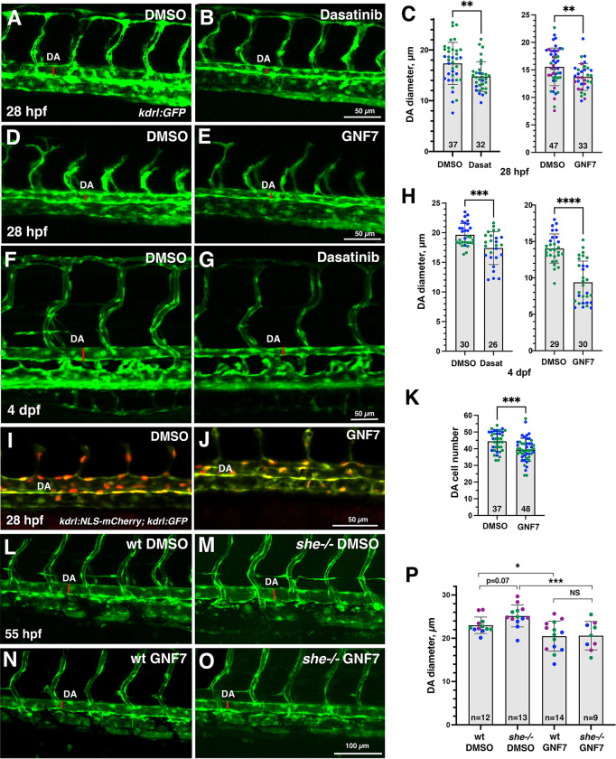
Fig 5. Inhibitors of Abl signaling reduce DA diameter in wild-type and she mutant embryos.
(A-E) Dorsal aorta diameter at 28 hpf is reduced in kdrl:GFP embryos treated with 5 μM Dasatinib or 1 μM GNF-7 compared to controls treated with 0.1% DMSO. (F-H) Embryos treated with 5 μM Dasatinib (F-H, left) or 1 μM GNF-7 (H, right) exhibit narrower DA at 4 dpf compared to controls treated with 1% DMSO (GNF-7) or 2% DMSO (Dasatinib treatments). (I-K) The number of cells in the DA is reduced in embryos at 28 hpf treated with 1 μM GNF-7 compared to control embryos treated with 0.1% DMSO. (L-O) GNF-7 treatment reverses DA enlargement in she mutant embryos. she+/-; kdrl:GFP adults were crossed to obtain she mutant embryos. Embryos were treated starting at 6 hpf with either 0.5 μM GNF-7 or 0.1% DMSO. Embryos were imaged at approximately 55 hpf and subsequently genotyped. DA measurements were performed blinded. Mid-trunk region is shown, anterior is to the left. Note the slightly wider DA (red line) in she-/- mutant embryos compared to wild-type (she+/+) siblings. DA is reduced in both wild-type and she mutant embryos treated with GNF-7. (P) Quantification of DA diameter in wild-type or she mutant embryos treated with GNF-7 or DMSO. In all graphs mean±SD is shown. Data points (shown in different colors) are combined from 2 (left graph C,H,K) or 3 (right graph C,P) independent experiments. Total number of embryos analyzed is shown at the bottom of each bar. *p<0.05, **p<0.01, ***p<0.001, ****p<0.0001, NS–not significant, Student’s t-test (C,H,K), or one-way ANOVA test, followed by multiple comparisons Fisher’s LSD (P).

