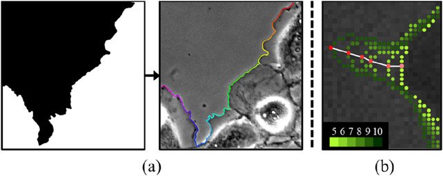
Figure 4.
(a) Extraction of contour points from the segmentation mask of one of the phase contrast live cell images [13]. Contour points are in sequential order as shown in color, from pink to red. (b) Labeling tracking points in 5x higher temporal resolution. The red point is tracked with correspondences shown in white lines. The color of the contour points changes from yellow-green to dark green as the frame number increases from 5 to 10.

